

"The true measure of a successful ablation is a pretty map." - Roy G. Biv. #proudtobeabbott #EPnerds #EPeeps #EPexplained AKA Aaron Stammer. Opinions mine
Joined December 2012
- Tweets649
- Following2.8K
- Followers1.5K
- Likes1.1K







Pinned Tweet
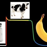
Now you’ll never forget the correct ion channel order for the cardiac action potential. #EPeeps


EP Nurse/Cardiac Mapper retweeted

3,822 doctors implanted left atrial appendage closure devices in 2025
My gosh
sciencedirect.com/science/ar…

While you’re waiting for Outlook to come back online, this is a refreshing use of your time!

Stop what you're doing an go read the comments on this tweet. Oh my word. This is the best use of X at the moment. 😂😂😂😂 @marklevinshow is a legend.
466
EP Nurse/Cardiac Mapper retweeted

Great resource for #EPeeps who want to return to EGM fundamentals.
This paper traces the EGM signal journey, explains the clinical consequences of morphological changes, and compares unipolar, bipolar, omnipolar, and multipolar EGMs.
Check it out: sciencedirect.com/science/ar…

EP Nurse/Cardiac Mapper retweeted

PVC localization has been one of the harder things to learn and teach as a cardiology fellow interested in EP.
So I built learnpvcs.com to help connect the 12-lead ECG with 3D cardiac anatomy.
You can explore different PVC origins, rotate and look inside the heart, and work through practice cases.
It's an early educational prototype, and I'd love feedback from fellows, electrophysiologists, and educators.
Which PVC origin do you find hardest to localize?
#CardioTwitter #EPeeps #MedEd @OpenAI @WvuCvFellows @wvudeptofmed

EP Nurse/Cardiac Mapper retweeted

I built NavRad Anatomy, a free tool with an AI Guide that makes it simpler to learn clinically important imaging anatomy.
To read scans, you need to know the anatomy. But there’s a lot to learn, and it’s hard to know what matters.
With this app, you can scroll through real scans with labels and colour-coded overlays, filter the structures you want to see, learn essential anatomy with key clinical pointers, or ask the AI Guide:
“Walk me through the liver segments” (see video)
“Teach me the segments of the internal carotid artery”
Or even: “Just show me the basics”
Then test yourself by asking it to quiz you.
Works on your phone or computer, so you can learn on the go.
See it in action 👇
Try it at anatomy.navigatingradiology.…, or click "Anatomy" in Navigating Radiology.
EP Nurse/Cardiac Mapper retweeted

I created LearnCoronary (learncoronary.com), an interactive tool designed to help general cardiology fellows and other early-career learners better understand coronary angiography and the spatial relationships between different angiographic views.
#cardiotwitter @OpenAI
EP Nurse/Cardiac Mapper retweeted

Efficiency Pro Tip ⌚
Use #RoboticEP automation features to help streamline procedure workflows and focus on other key aspects of the procedure.
Learn more about enhancing your lab with #RoboticEP at roboticep.com/
EP Nurse/Cardiac Mapper retweeted

Taquicardia por Reentrada Nodal AV.
⚡️🫀 @hrs_journal Review2026 👌🏻
🔶️ La TRNAV es la taquicardia regular más frecuente. El término “nodal” podría ser anatómicamente impreciso: el circuito utiliza estructuras por encima, alrededor y por debajo del nodo AV, por lo que se ha propuesto llamarla reentrada de la unión AV. 🤔💓
🔸 TRNAV típica (slow-fast): conducción anterógrada por la vía lenta y retrógrada por la rápida. En el EKG, la P retrógrada suele quedar oculta dentro del QRS o aparecer como una pequeña pseudo-r′ terminal. En el EEF: AH/HA >1 y VA ≤60 ms.
🔸 TRNAV atípica: la P retrógrada es visible antes del siguiente QRS → RP > PR, habitualmente negativa o poco profunda en II, III y aVF y positiva en V1. Representa ≈6% de las TRNAV. La clasificación clásica fast-slow vs slow-slow tiene poca relevancia práctica; Katritsis propone simplificarla en típica vs atípica, utilizando principalmente HA y VA: típica HA ≤70 ms / VA ≤60 ms y atípica HA >70 ms / VA >60 ms.
🔶️ Tto farmacológico: en el episodio agudo se utilizan primero maniobras vagales y adenosina (menos eficaces en TRNAV que en TRAV). Si fracasan y aparece hipotensión, está indicada la cardioversión eléctrica sincronizada. 💊⚡️
🔶️ La ablación alcanza ≈97% de éxito, recurrencia ≈1.3–4% y una tasa global de complicaciones ≈0.3% 🔌🔥
📝🆓️⤵️
doi.org/10.1016/j.hrthm.2025…
t.me/medicinainternaconsoque…


EP Nurse/Cardiac Mapper retweeted

High-fidelity correlation between predictive E-field and final lines #Volt

EP Nurse/Cardiac Mapper retweeted
🫀⚡ SMART-WPW: nuevo algoritmo EKG para localizar vías accesorias.
🔶️ Desarrollado y validado en 260 pacientes, incluidos 76 menores de 18 años, y permite identificar 12 sitios anatómicos de ablación mediante una representación tipo “reloj” de los anillos tricuspídeo y mitral. El algoritmo se basa en 3 pasos:
1️⃣ Polaridad de la onda delta en V1 → derecha vs izquierda. ↔️
▪️Delta negativa en V1 → vía derecha (anillo tricuspídeo). Un patrón qS orienta hacia vía septal, mientras que rS favorece pared libre derecha.
▪️Delta positiva en V1 → vía izquierda (anillo mitral); algunas vías izquierdas posteriores/laterales pueden ser isoeléctricas.
2️⃣ Si la vía es derecha, mirar V3 → septal vs pared libre. 🤔🔎
➕ R prominente en V3 → septal.
➖ S prominente/rS → pared libre derecha.
3️⃣ Polaridad de la delta en II, III y aVF → posición final (anterior o posterior).
Se cuenta cuántas derivaciones inferiores presentan delta negativa (0–3). A medida que aumenta la negatividad inferior, la vía se localiza progresivamente más posterior; una delta más positiva indica una posición más anterior.
📊 Rendimiento: localizó correctamente la vía en 253/260 pacientes (97%), con sensibilidad 96%, especificidad 100%, VPP 95% y VPN 100%. Superó a Pambrun (97% vs 90%) y Arruda (97% vs 73%). En pacientes pediátricos mantuvo 97% de precisión, frente a 62% con Arruda. 👌🏻✔️
📝🆓️⤵️
doi.org/10.1016/j.hrthm.2025…
t.me/medicinainternaconsoque…


EP Nurse/Cardiac Mapper retweeted

🔥 Hot off the press: We identified how and where AF begins.
I am excited to share this work.
Since the discovery of PV triggers more than 25 years ago, AF ablation has evolved into an empiric therapy centered on PVI. Yet, we have never been able to identify the arrhythmogenic substrate that determines why a premature beat initiates AF at one site and not another.
We believe this study changes that.
In this mechanistic study, we found that AF initiation arose predominantly through reentry originating from a small number of spatially discrete regions with steep repolarization gradients. Their distribution was unique to each patient, and they accounted for more than 92% of AF initiation episodes.
Importantly, the arrhythmogenic substrate was not defined by low voltage, and while conduction abnormalities were often present, they appeared to play a secondary role.
Its patient-specific distribution may help explain the controversy surrounding posterior wall isolation: the posterior wall harbored the substrate in only approximately 30% of patients. Importantly, in redo patients with durable PVI, right-atrial substrate (which is often beyond standard mapping strategies) was identified in 30%.
Most encouragingly, in an exploratory cohort of 24 patients with recurrent AF and durable PVI (50% PerAF), targeted elimination of these sites achieved 87.5% single-procedure freedom from any atrial tachyarrhythmia over a median of 489 days, without antiarrhythmic drugs.
The thoughtful accompanying editorial by Jon Kalman highlights the significance of this discovery and its central translational challenge of bringing it into real-world clinical practice.
But stay tuned! Technology is under development to transform this discovery into a rapid, automated functional mapping.
Current electroanatomic mapping relies largely on voltage amplitude and activation timing. We need to move beyond these measures and map how tissue actually functions - how it conducts, repolarizes, and responds to premature stimulation. This can add the critical dimension that has long been missing from substrate mapping and may reveal what truly makes cardiac tissue arrhythmogenic.
To me, this is the beauty of physiology: there are no black boxes. We can identify the sites that reproducibly initiate the arrhythmia, define their EP properties, eliminate them with ablation, and confirm that arrhythmia can no longer be initiated.
The next steps are prospective multicenter validation and technological development. Together, they may move AF ablation beyond empiric therapy toward truly individualized, mechanism-guided therapy.
📄 Paper in @JACCJournals:
doi.org/10.1016/j.jacep.2026…
📝 Editorial:
doi.org/10.1016/j.jacep.2026…
#JACCCEP #AtrialFibrillation #CardiacElectrophysiology #CatheterAblation #EPeeps

EP Nurse/Cardiac Mapper retweeted

The EP Fellow Bootcamp has begun! 🚀
An insightful educational session with Dr. @DJ_Lakkireddy is underway, empowering the next generation of EP fellows with expert knowledge and practical insights.
#EPFellowBootcamp #Electrophysiology #Cardiology #MedicalEducation


EP Nurse/Cardiac Mapper retweeted

QRS AXIS IN THE FRONTAL PLANE DURING CARDIAC PACING ❗
Today I made a thread about magnet 🧲 ECG with RV apex pacing.
(From: Cardiac Pacemakers Step by Step An Illustrated Guide by S. Serge Barold, Roland X. Stroobandt, Alfons F. Sinnaeve)

EP Nurse/Cardiac Mapper retweeted

Surprise spontaneous ORT using a concealed left lateral pathway on a younger patient (early 40s) scheduled for PFA AF ablation (history of persistent AF). Should we still proceed with the PVI? #EPeeps

EP Nurse/Cardiac Mapper retweeted

The coronary arteries fill with blood during:
A. atrial systole.
B. atrial diastole.
C. ventricular systole.
D. ventricular diastole.

EP Nurse/Cardiac Mapper retweeted
Pinwheel 🎡 flutter in the RSPV before starting PFA PVI. Patient presented in this rhythm and ECGs going way back show only this AFL, sometimes 2:1 and sometimes Wenckebach. Do you isolate the RSPV and call it good, isolate both RPVs, or perform the full PVI as planned? #EPeeps
EP Nurse/Cardiac Mapper retweeted

My new favourite entrance to a cardiac department!! (Created by Angela Palmer) #echofirst #cardiotwitter #whyCMR
EP Nurse/Cardiac Mapper retweeted

Supraventricular tachycardia (Source Unknown) #MedEd