

Become a better endoscopist with EndoCollab, the world's most comprehensive endoscopy community. Join us: https://nitter.cf/t.co/EkRtunpQyB
🌎
Joined February 2021
- Tweets10.2K
- Following2.9K
- Followers31.2K
- Likes21.2K




EndoCollab™ | GI Endoscopy Community retweeted

About 8 mm: a ring-like duodenal stenosis the scope could not pass. Balloon over a guidewire, inflated with water under direct view, watching the mucosal rips. After: breaks even and not deep, and the scope passed easily.
By Klaus Mönkemüller, MD, PhD
EndoCollab™ | GI Endoscopy Community retweeted
Same patient. A: standard view, mucosa looks "normal". B: water immersion shows erythema, edema and tiny angiodysplasias.
Portal hypertensive duodenopathy is often missed. Water immersion and NBI make it more evident.
By Jay Bapaye, MD and Klaus Mönkemüller, MD, PhD
EndoCollab Quiz
Gastric polyposis on EGD. Colonoscopy was normal and there were no duodenal polyps.
What is the diagnosis?
a. Peutz-Jeghers syndrome
b. FAP
c. Juvenile polyposis syndrome
d. Cowden syndrome
e. MAP
f. GAPPS
Vote in the reply to see the answer.

Answer in our Telegram quiz. Vote to see it:
t.me/endocollab/86

42M, acute abdomen, cholestatic liver tests, dilated bile duct.
Massive papillary edema (explains the duct), purplish duodenum.
Dx: IgA vasculitis (Henoch-Schönlein purpura). Any age. Key finding: palpable purpura.
On your differential?
Full lecture on EndoCollab: endocollab.com/blogs/full-co…

Five endoscopy classifications on one page: Prague, Hill, Forrest, Paris and EREFS.
Barrett's extent, flap valve, ulcer stigmata, lesion shape and EoE findings.
Shared grading means the next endoscopist reads your report the way you meant it.

The Paris classification applied to colon polyps, from the Train Your Eye course: endocollab.com/blogs/member-…

Acute pancreatitis has a longer cause list than gallstones and alcohol.
Read a few entries with caution: sphincter of Oddi dysfunction, pancreas divisum and gallbladder polyps are contested or weakly supported causes.
Before calling it idiopathic, check triglycerides and calcium.
Adapted from United European Gastroenterology Journal, 2020.

Spurting vessel in D2. No ulcer around it.
37F, hematochezia, CKD, diabetes. That nipple is a Dieulafoy. One through-the-scope clip. Field dry.
Clip first or inject first when the vessel is already in view?

EndoCollab™ | GI Endoscopy Community retweeted
Hemorrhagic shock on clopidogrel. CTA: mid-jejunal bleed. Enteroscopy: jejunal diverticulum with ulcer, treated with clips. Case by Klaus Mönkemüller, MD, PhD.


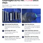
Esophageal SEMS, the 2 + 2 rule:
stent = stricture + 2 cm above + 2 cm below.
3 cm stricture -> 7 cm stent. Not too long.
The proximal string lets you pull it back to reposition.
Skin marker or internal clip?

Full lecture on EndoCollab: endocollab.com/blogs/full-co…

EndoCollab™ | GI Endoscopy Community retweeted
Two red, edematous antrums. A: EBV lymphoproliferative disorder that mimicked GAVE. B: nodular GAVE, pseudo-polypoid.
EBV can mimic GAVE. Biopsy tells them apart. Keep EBV in mind when suspected GAVE looks unusually edematous.
By Klaus Mönkemüller, MD, PhD and Jay Bapaye, MD
Exploring the colon with water:
1. Better navigation through natural kinks or a stenosis
2. Helps ileum intubation
3. Clears debris so you can study the polyp surface
4. A tool for underwater resection
Do you use water routinely?
History of Meckel's Diverticulum
- The first time it was described in 1809
- Meckel's rule of 2's
- A nice graph showing potential lengths
- Endoscopic spectrum of Meckel's diverticulum
EndoCollab Quiz
Pseudopolyposis of the stomach
Patient with malabsorption, alopecia, brittle nails
What is your diagnosis?
Reply below with your answer.


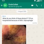
From the EndoCollab WhatsApp group: a 53-year-old woman hospitalized with DKA, with odynophagia. A member asked: "Just candidiasis?"
Prof. Klaus Mönkemüller: "This is severe erosive esophagitis associated with DKA."
His full answer is in the second image.


