
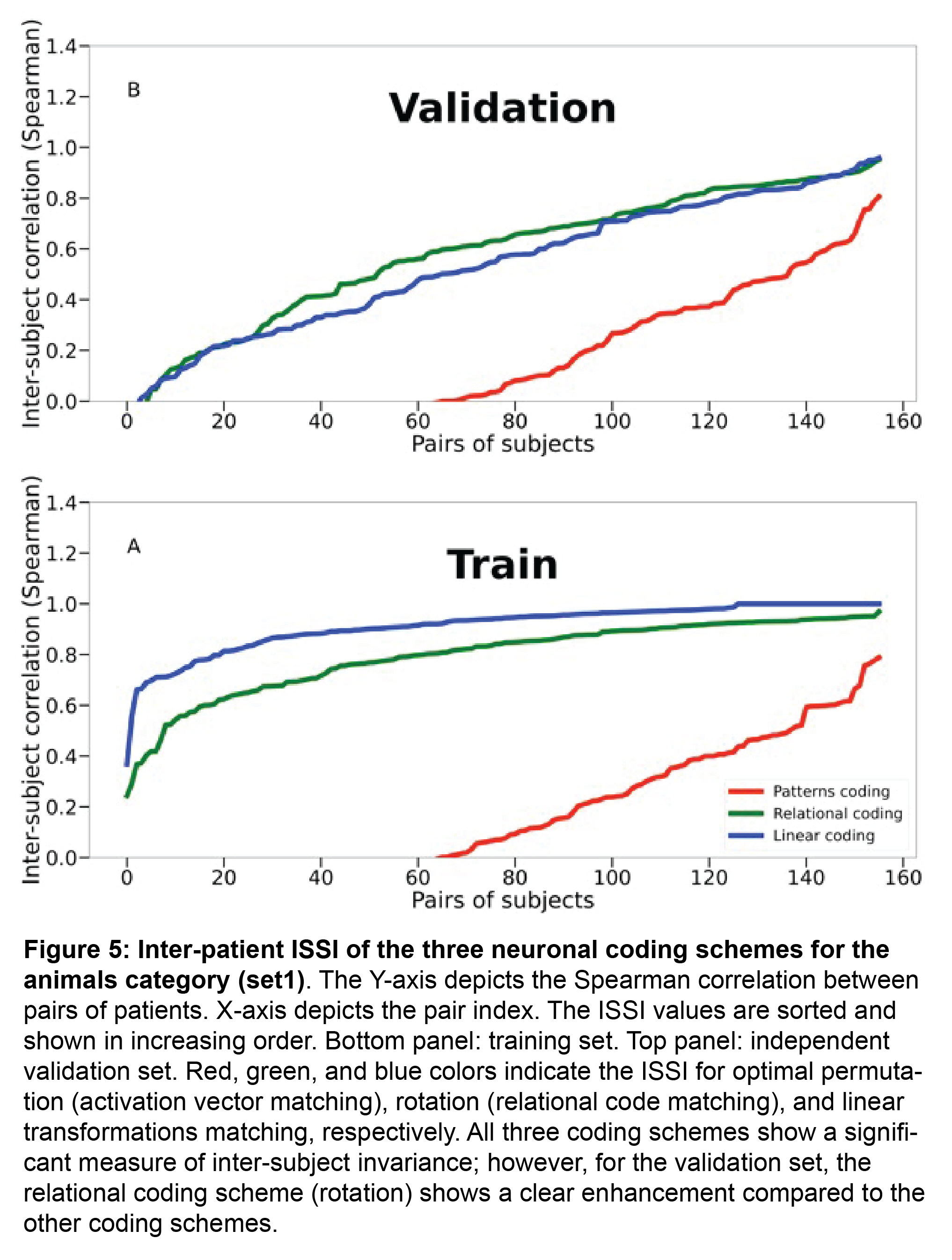
However, our paper refutes this notion by showing that what remains most stable across individuals’ brains are the similarity structures -- the set of similarities and dissimilarities between activation patterns of each image to other images from the same category (see figure).

As can be seen, the receptive field structure of entorhinal neurons resembles the basis functions of JPEG, suggesting a form of lossy representation of space in the entorhinal cortex. (4/4)

Our results show that what remains invariant across brains is not the activation patterns (population vectors), commonly considered the “work horse” of visual representations, but rather the similarity structure (relational coding) which emerges between these patterns (see fig)
5/6 These results suggest that relational coding, rather than the absolute population vector amplitudes, constitute the underlying mechanism of perceptual stability in high-order visual regions.

4/6 what remains stable over time is the relative activations of individual neuronal groups for different images, i.e. the activation profiles, and the set of similarity distances between these different activity patterns.

3/6 In our study we demonstrate, using iEEG recordings in patients, that although the average activation magnitude in high order visual areas drops by ~70% when patients view a stationary visual image for an extended time period (> 1s),

1/6 What keeps our visual perception stable across sustained time durations, despite a dramatic reduction in neuronal activity?
We are pleased to share our work that has just been published in Cell Reports sciencedirect.com/science/ar… suggesting a plausible answer to this question.

4/5 The stability was evident in visually-responsive contacts in high order occipito-temporal cortex but not in frontal cortex- a finding relevant to general theories of conscious perception.

Finally, we tackle the critical open question of which regions in the cortex interact with these retrieval-related ripples. We found a wide-spread dialogue between the hippocampus and the neocortex, with strongest peri-ripple activity within the default mode network (DMN). 9/10

