

Chief of Radiology| 4x Clinical Fellowships| MSK & Sport Med LA, 🇺🇸|Abdomen Singapore, 🇸🇬|Head & Neck London,🇬🇧|Paediatrics, Ottawa 🇨🇦
Canada
Joined April 2022
- Tweets172
- Following41
- Followers1.2K
- Likes23





🧠 Being a Clinical Radiologist
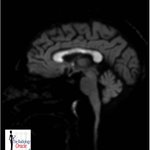
👨⚕️ 40-year-old man with Confusion + Altered Consciousness
There was no prior physician evaluation as patient presented initially via a proxy.
An MRI brain was advised based on the presenting complaints.


💡 Discussion:
The distribution was unusual enough to prompt a crucial clinical question: “Is there a history of chronic alcohol use?”
The history was initially denied. We asked the patient's wife. She disclosed long-standing alcoholism.
🧠 QUIZ TIME!
❓ Recommendations you would absolutely NOT make here ?
Ans with your Best Memes 🤓
#MedEd #Neurology #Radiology
#RadRes #Neurosurgery
@_the_SRT @spinacademics @thecortexclub


1
234
📖 Radiology textbooks: “CT and MRI have taken over.”
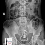
👨🏫 Examiners: “Anyway… identify this barium meal follow-through.”
Some things age, but exam questions don’t.
🎯 High-yield. Old-school. Guaranteed to show up when you least expect it.
#MedStudent
#MedEd

1
8
727
🚨 1 in a Million Case | Arterio-Colic Fistula 🚨
CT reveals an extraordinary finding; a thin channel communicating directly from the LEFT external iliac artery to sigmoid colon producing a hyperdense intraluminal appearance.
#RadTwitter #MedicalImaging #FOAMed #MedEd

2
301
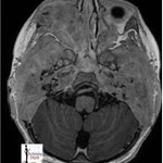
18-month-old boy with bilateral post-auricular swelling 👶🦴
Clinical presentation:
🔹 Otorrhea with mastoiditis
🔹 Poor response to sounds
🔹 Gradual hearing loss reported by parents
#FOAMRad
#MedEd
@_the_SRT @ESHNRSociety @spinacademics


1
4
351
💡 Diagnosis:
Skull Base Langerhans Cell Histiocytosis (LCH)
Temporal bone LCH in children can closely mimic aggressive otomastoiditis or neoplastic pathology. Bilateral destructive temporal bone lesions with hearing symptoms should always prompt consideration of LCH.
1
156
⚠️ Imaging pearl:
Marked osseous destruction out of proportion to the apparent infectious process.
What would be your top differential diagnosis before biopsy?👇
📌 Please Consider Sharing to Spread the Word !
instagram.com/radiologyoracl…
167
🧠 Rare Pelvic Case | Radiology Spotlight
A well-encapsulated lesion with innumerable internal cysts is seen in the RIGHT adnexal region displacing the urinary bladder without perifocal infiltration.
👉 Classic imaging appearance raises strong suspicion for Hydatid Disease


📌 Diagnosis
🔎 Pelvic Hydatid Cyst
🩺 Post-op: Primary Giant RIGHT Ovarian Hydatid Cyst
💡 Why is this interesting?
Hydatid cysts (caused by Echinococcus granulosus) most commonly involve the liver and lungs.
👉 Ovarian involvement is extremely rare !
With immense pride and sincere gratitude, I wish to share that I have been conferred the Dr. Kishor Taori Gold Medal at the IRIA Annual National Conference, Hyderabad 2026.
This prestigious award was presented during the IRIA Oration Session, along with a Medal and Citation.


2
4
603
TheRadiologyOracle retweeted

Cafe Roentgen REF Wednesday Webinar #927- Hidden Areas of Chest and Abdomen on Plain Radiographs - @theradoracle, Indore
When: 19 Nov, 7-8 pm IST
Link: us02web.zoom.us/j/8566622006…
Webinar ID: 85666220065
Passcode: REFCAFE
#radres #radtwitter @FOAMrad @RadioGyan @rsna @myESR

20 Day Old Baby with Fever and Seizures
Diagnosis in Thread
#FOAMed #FOAMRad
#MedTwitter
#Neurosurgery
#Medstudent
#Radiology


On T2, FLAIR, T1 C+ and DWI Images:
There are 2 distinct thick walled rim enhancing areas with heterogeneous morphology within both frontal lobes. Significant restriction is seen on diffusion acquisition in keeping with liquefied contents.