
AP/CP Pathologist @KingsCollegeLon Trained @MayoClinicPath @Yalepathology @NU_pathology | Digital Pathology | AI in Medicine | Sharing real diagnostic cases
Joined October 2025
- Tweets531
- Following7.5K
- Followers920
- Likes7K




A suprarenal mass. Radiology says adrenocortical carcinoma.
The core shows spindle cells in fascicles. Inhibin: negative.
A) Leiomyosarcoma
B) Adrenocortical carcinoma
C) GIST
Which stain would you order next? Answer tomorrow at noon.
#PathTwitter #SoftTissuePath

'Positive' was negative.
Answer: B. Invasive lobular carcinoma.
The E-cadherin was read as positive. Look closer:
1. Discohesive, single-file cells on H&E
2. E-cadherin dot-like and fragmented, not membranous
3. Beta-catenin: membranous staining lost
A positive stain in the wrong pattern is a negative result.
#Pathology #BreastPathology

🍁 BACK IN MICHIGAN — WHERE MY JOURNEY BEGAN!
Beautiful fall colors. Wonderful memories. My early education. My home state. ❤️
And this visit comes with another BIG excitement!
📖 MY BOOK IS COMING!
LAB BLACK BOXES 🔬
The manuscript is submitted. Hopefully releasing NEXT WEEK, Insha’Allah!
We rely on laboratory results for countless clinical decisions.
But how many physicians truly understand what happens INSIDE the laboratory’s digital systems?
The analyzers.
The middleware.
The LIS.
The interfaces.
The hidden databases.
The data behind every result.
And what happens when these systems fail?
These are the BLACK BOXES of laboratory medicine.
My mission is simple:
OPEN THE BOXES. MAKE THE COMPLEX SIMPLE. UNLOCK THE DATA.
For physicians, laboratory professionals, researchers, and anyone exploring AI in healthcare.
No programming experience required!
I’ll do my best to keep the book affordable and accessible.
🍁 From Michigan memories to a new educational journey.
LAB BLACK BOXES — COMING SOON!
#LabBlackBoxes #LaboratoryMedicine #HealthcareAI #PatientSafety
🤖 Made with AI


10 YEARS OLD. 100 KILOGRAMS. CHILD OR ADULT PATIENT?
Biologically, a child. Medically, potentially facing complications we once associated mainly with adults.
Recently, we encountered a 24-year-old with esophageal adenocarcinoma.
It made me wonder: What if severe obesity and reflux had been present since childhood?
We cannot know that patient’s earlier history. But consider the thousands of children living with severe obesity today.
WHO IS RESPONSIBLE FOR THEIR CARE?
Pediatricians? Adult physicians? Endocrinologists? Gastroenterologists? Bariatric specialists?
What happens at 14, 16 or 18?
Who decides when care transfers?
Can pediatric wards accommodate their physical and clinical needs?
WHO provides age-specific obesity definitions. Pediatric treatment guidelines exist.
But guidelines alone do not guarantee coordinated care.
What we urgently need are clearly defined responsibilities, specialist pathways, age-appropriate facilities and a formal transition to adult care.
A child does not become an adult because they weigh 100 kilograms.
And they should not be lost between pediatric and adult healthcare systems.
This is not simply an obesity problem.
It is a healthcare-system design problem.
#ChildhoodObesity #Pediatrics #PublicHealth #HealthcarePolicy #CancerPrevention


Extracellular mucin in a 2 cm colon polyp. 57-year-old man.
Has it crossed the line into mucinous adenocarcinoma?
Look at where the mucin sits:
It stays with the dysplastic glands.
No desmoplasia. No single infiltrating cells. Nothing beyond the muscularis mucosae.
Final: tubulovillous adenoma with high-grade dysplasia and mucinous differentiation.
It is not how much mucin you see.
It is where it is, and how it behaves.
#PathTwitter #GIPath


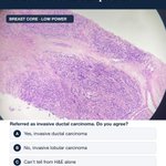
Breast core. Referred as invasive ductal carcinoma.
E-cadherin was reported positive.
A) Agree: invasive ductal carcinoma
B) Disagree: invasive lobular carcinoma
C) Can't tell from H&E alone
Answer tomorrow at noon.
#Pathology #BreastPathology

Crowded glands, a quarter of the cervix.
Answer: B. Benign. Focal atypical endocervical glandular hyperplasia.
Why not adenocarcinoma:
1. Focal
2. No deep stromal invasion
3. No desmoplastic response
4. Mild cytologic atypia only
Cervical cancer diagnosis is not just about spotting cancer. It is about not creating one.
#PathTwitter #GynPath

Our genes have not changed in a generation.
Our world has.
In one generation, the Gulf has seen:
Rapid urbanisation.
A dramatic change in diet.
Vitamin D deficiency despite abundant sun.
Rising obesity.
All on a background of high consanguinity.
Every one of these sits on the list of suspects for autoimmune disease.
So we may be living the hygiene hypothesis in fast-forward.
I say hypothesis deliberately.
Regional registries and good lab data could answer it.
Too clean, too fast?
What would you measure first?

A low-grade endometrioid carcinoma. 22 lymph nodes negative. Ki-67 relatively low.
Yet the patient had widespread metastatic disease.
This is p53 from the low-grade areas. Brown nuclei everywhere.
Wild-type or mutant?
Make your call before you open the replies.
#Pathology #GynPath
REPLY (post as a reply, 18:00 UTC under W4-P19)
Answer: wild-type.
Most of this tumor showed the expected heterogeneous wild-type pattern. Only one geographically distinct, higher-grade focus raised the subclone question.
Variable intensity plus scattered negative nuclei = wild-type.
Strong, diffuse, near-uniform staining = abnormal.
Complete null with intact controls = abnormal.
Pattern > %. Always.

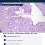
56-year-old woman. Cervical biopsy.
Crowded, irregular glands. But focal, with only mild atypia.
A) Adenocarcinoma in situ
B) Benign glandular hyperplasia
C) Invasive adenocarcinoma
Reply with your call and one reason. The answer lands at noon.
#PathTwitter #GynPath

Not a myxoma. Not in a vessel.
Answer: C. Myxoid endometriosis, decidualized and gland-poor.
Two traps in one slide:
1. A myxoid, gland-poor nodule that reads like a myxoma
2. A space that looks like a vessel, with no endothelial lining
Immunohistochemistry confirmed endometrial stromal differentiation.
Not every vascular-looking space is a vessel.
#Pathology #GynPath

Ki-67 ABOVE 40%. MITOSES BELOW 5. IMMUNOSTAINS THAT REFUSED TO COOPERATE!
What kind of juvenile granulosa cell tumor is THIS?
A 34-year-old woman. An 8 cm ovarian tumor. A diagnostic puzzle!
Morphology favored juvenile granulosa cell tumor (JGCT).
But the immunohistochemistry told a very strange story!
• Inhibin: Strongly positive in scattered peripheral/theca-like cells, but most solid tumor nodules were NEGATIVE!
• Calretinin: Similar unusual peripheral and stromal-associated staining!
• WT1: Heterogeneous nuclear positivity.
• Ki-67: 40–50% in hotspots!
• Mitotic count: Only 3–4 per 10 HPF.
HOW CAN SO MANY CELLS BE PROLIFERATING WHEN SO FEW ARE DIVIDING?
Possible explanations:
Ki-67 marks cells throughout the active cell cycle—not just mitosis.
Proliferative hotspots may differ from areas used for mitotic counting.
Tumor heterogeneity, cell-cycle kinetics, and counting methodology may contribute.
And the unusual inhibin/calretinin pattern? Possibly heterogeneous sex cord differentiation or a distinct luteinized/theca-like cell population.
THE PATHOLOGY TWIST:
High Ki-67 does not automatically mean a poorly differentiated tumor.
Focal inhibin positivity does not exclude granulosa cell tumor.
And unusual immunostains do not automatically establish Sertoli–Leydig differentiation.
Despite all these contradictions, the predominant follicular and lobulated morphology still favored JGCT.
PATHOLOGY PEARL:
The microscope shows what cells look like.
Ki-67 shows how many are cycling.
Mitoses show how many are visibly dividing.
They are related—but they are NOT the same measurement!
When morphology, immunostains, and proliferation disagree, which finding would you trust most?
#Pathology #JuvenileGranulosaCellTumor #Ki67 #Immunohistochemistry #GynecologicPathology


When can a child with coeliac disease skip the biopsy?
Under the ESPGHAN 2020 guideline, when:
tTG-IgA is at least 10 times the upper limit of normal,
and EMA-IgA is positive in a second blood sample,
and the family agrees.
That puts a lot of weight on one number.
So the lab has to earn it:
The assay must measure reliably up to and beyond 10 times the limit.
The report must make that multiple clear.
When a lab number replaces a biopsy, the number has to be right.
Does your report show the multiple of the upper limit?

Someone asked under my thyroid capsule post:
"Can follicular carcinoma present with invasion and a thin, smooth capsule, or must invasion be present in addition to a thick, irregular capsule?"
The capsule's look is not the criterion. Invasion is.
A capsule can be thick, fibrotic, irregular, frankly frightening, and the lesion can still be benign.
Size alone does not make it cancer.
Cytologic atypia alone does not make it follicular carcinoma.
What matters: convincing capsular and/or vascular invasion. That is why we examine the interface between tumor and capsule so carefully.
Keep the questions coming. The best ones become posts.
#PathTwitter #ThyroidPath


Abdominal wall nodule.
Myxoid. No atypia. No destructive growth. And it seems to sit inside a vessel.
A) Myxoma
B) Intravascular tumor
C) Endometriosis
Which stain would you order first? Answer tomorrow at noon.
#Pathology #GynPath

Lots of fungus. No invasion.
Answer: B. Allergic fungal rhinosinusitis.
1. Fungi trapped in allergic mucin, not in tissue, vessels or bone
2. Vessels: reactive, driven by chronic allergy, not angioinvasion
3. Crystals: Charcot-Leyden, the footprint of eosinophils
The fungus sits in mucin. The reaction is allergic, not invasive.
Abundant fungus does not equal invasion.
Misread it and the patient gets antifungals they do not need.
#PathTwitter #HeadAndNeckPath

Mild stomach. Dramatic esophagus. 34-year-old man.
A circumferential ulcer in the distal esophagus, and a gastric biopsy that looked almost quiet.
How I worked it:
1. Rule out viral cytopathic effect
2. Rule out fungal organisms
3. Then call severe reflux injury
4. And check the antrum: H. pylori gastritis
Sometimes the stomach biopsy whispers.
And the esophagus shouts the diagnosis.
#Pathology #GIPath


🔬 Myxoid Change + Adenosis: A Histological Trap!
A fibroepithelial breast lesion can look surprisingly complex!
🧬 Myxoid stromal change creates a loose, sometimes alarming appearance.
🔍 Prominent adenosis makes the lesion look crowded and architecturally busy—but highlights its benign epithelial component.
⚠️ Don’t mistake myxoid change for stromal overgrowth!
The real clues to phyllodes tumour? Stromal cellularity, subepithelial condensation and exaggerated leaf-like architecture.
💡 A busy-looking lesion isn’t necessarily an aggressive lesion!
#BreastPathology #Fibroadenoma #PhyllodesTumor


57-year-old woman. Nasal polyps.
Abundant fungus. Striking vessels. Eosinophils. Crystals.
Invasive fungal sinusitis is an emergency. Is this it?
A) Invasive fungal sinusitis
B) Allergic fungal rhinosinusitis
C) Fungal infection: start antifungals
The answer, with the clue circled, lands at noon.
#PathTwitter #HeadAndNeckPath

Cysts at low power. Trophoblast at high power.
Answer: B. Placental site trophoblastic tumor.
Not a mole with a tumor on top. Moles and PSTT come from different trophoblast lineages.
What it showed:
1. Sheets and cords of intermediate trophoblast
2. Vascular wall invasion and replacement
3. Abundant fibrinoid
The "cysts" were vascular and fibrinoid remodeling spaces, not molar villi.
Know the trophoblast, and the diagnosis follows.
#Pathology #GynPath
