aquifer (@jppathpd) profile picture

Pathologist #PathKeys

Posts = my own opinion
Joined November 2022
Photo by aquifer (@jppathpd)Photo by aquifer (@jppathpd)Photo by aquifer (@jppathpd)Photo by aquifer (@jppathpd)Photo by aquifer (@jppathpd)Photo by aquifer (@jppathpd)Photo by aquifer (@jppathpd)Photo by aquifer (@jppathpd)Photo by aquifer (@jppathpd)Photo by aquifer (@jppathpd)
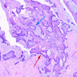
Dx? mandible HE #Pathkeys 2025-1p-2 (etc)
1
2
12
739
⭕Ameloblastoma (follicular and plexiform patterns) 🔬Epithelial islands with peripheral palisading of columnar cells and central stellate reticulum-like cells in a fibrous stroma ☢Well-defined radiolucency around the crowns of impacted molars with partial cortical resorption
1
5
71
Image (pathology, panoramic radiograph): Gao X, et al. Front Oncol 2026;16:1801359 (CC BY 4.0) Ref: Gao X, et al. Front Oncol 2026;16:1801359
57
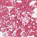
Dx? pleura STAT6 #Pathkeys
2
1
14
646
🧬NAB2-STAT6 fusion (intrachromosomal rearrangement on 12q) ☢️Well-circumscribed pleural-based mass, mostly attached to visceral pleura; may grow large enough to fill a hemithorax and displace the mediastinum
1
2
82
Image (pathology): Paraschiv A, et al. Reports 2026;9:229 (CC BY 4.0) Image (CT): Paraschiv A, et al. Reports 2026;9:229 (CC BY 4.0)
55
Dx? adrenal chromogranin A #Pathkeys
1
1
10
568
🧬At least 30% hereditary: SDH genes (most common), NF1, VHL, RET, MAX, EPAS1, FH; SDHB IHC screens for SDH loss ☢️Marked T2 hyperintensity ("light bulb sign"); no signal drop on opposed-phase MRI, unlike lipid-rich adenoma
1
64
Image (pathology): Zhang J, et al. Front Oncol 2026;16:1770384 (CC BY 4.0) Image (MRI): Tamayo Y, et al. Abdom Radiol 2026;51:3004-9 (CC BY 4.0)
35
Dx? kidney HE #Pathkeys
1
2
14
781
⭕oncocytoma 🔬Nests and tubules of cells with abundant granular eosinophilic cytoplasm and small round regular nuclei, floating in edematous stroma 🧪c-kit+; CK7- 🧬Loss of chromosome 1, 11q13 rearrangement, deletion of chromosome 14 ☢️Central scar may be seen on CT
1
5
170
Image (pathology): Pagano D, et al. World J Surg Oncol 2018;16:123 (CC BY 4.0) Ref: Goldblum JR, et al. Rosai and Ackerman's Surgical Pathology, 11th ed. Elsevier; 2018
1
110
To everyone who follows me: thank you, as always, for reading, liking, and reposting. This account posts one case a day, drawn mainly from diseases that have appeared on Japan's pathology board examination (every image cited comes from CC BY papers only).
1
1
6
362
My aim is the sharpest possible diagnostic skill — reading an H&E image alone and "seeing through" it to what lies beyond the disease and to its genetics — and I will keep working toward it. Thank you all for your continued support.
4
59
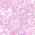
Dx? kidney Pt with dialysis #Pathkeys
1
6
21
839
⭕ Acquired cystic disease-associated RCC 🔬 Arises in ACKD. Eosinophilic cells with prominent nucleoli and cytoplasmic vacuoles giving a sieve-like pattern; intratumoral calcium oxalate crystals. ☢️ small atrophic kidneys studded with cysts + heterogeneously enhancing solid mass
1
1
6
310
Image (pathology): Zhou L, et al. Front Oncol 2024;14:1349610 (CC BY 4.0) Image (CT): Yang Y, et al. Front Oncol 2023;13:1187495 (CC BY 4.0) Ref: El-Zaatari ZM, Truong LD. Biomedicines 2022;10:657 Ref: Yang Y, et al. Front Oncol 2023;13:1187495
2
105
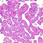
Dx? kidney GATA3 #Pathkeys
2
18
70
2,899
🧬KRAS mutation in most cases (~80–90%), clustered in exon 2 codon 12 (mainly G12V); no trisomy 7/17. Indolent behavior ☢️Small, round, well-defined renal mass with high attenuation on pre-contrast CT (arrow)
1
2
319
Image (pathology): Mohanty SK, et al. Virchows Arch 2024;485:829 (CC BY 4.0) Image (CT): Lee HJ, et al. Jpn J Radiol 2024;42:1458 (CC BY 4.0) Ref: Marletta S, et al. Virchows Arch 2026;489:631 Al-Obaidy KI, et al. Mod Pathol 2022;35:1279
234