

Médico Radiólogo con experiencia en Tomografia y Resonancia. Especialidad en imagen cardiovascular avanzada (resonancia cardíaca y tomografía cardíaca).
Caracas, Venezuela
Joined June 2013
- Tweets3.4K
- Following1.6K
- Followers931
- Likes6.4K

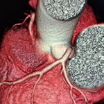





Cardiopatía isquémica crónica
Estudio para valorar viabilidad
#UrológicoSanRomán


Dra. Luisa Vera retweeted

Metatarsalgia: evaluación paso a paso
🔍 Este #PiperSERAM evalúa la utilidad y los hallazgos de las diferentes pruebas de imagen en el diagnóstico de la metatarsalgia, desde la radiografía simple hasta la RM, pasando por la ecografía y la TC, con correlación entre técnicas y un amplio espectro de entidades causales
Accede aquí 🔗f.mtr.cool/qsmo2yh0je


Dra. Luisa Vera retweeted

1/Have MULTIPLE questions about the new criteria for MULTIPLE sclerosis?
Do you know the NEW McDonald criteria for multiple sclerosis?
This month’s @theAJNR SCANtastic has the lastest you need to know!
ajnr.org/content/47/9/2454


Dra. Luisa Vera retweeted

⚠️COVID-19⚠️
🔴Síndrome del Corazón Roto🔴
➡️Miocardiopatia por stress debido a la liberación hormonas cómo epinefrina y norepinefrina entre otras
➡️Estudio de la Cleveland Clinc en Ohio, USA y publicado en JAMA con 1,900 pacientes con dolor torácico en la época de COVID

Dra. Luisa Vera retweeted

❗️No existe un límite para grosor del miocardio apical para diagnosticar una miocardiopatia hipertrofica apical
🔎La idea : definir un grosor máximo del apex en personas sanas
📍un grosor máximo apical de 5.2 a 5.6 mm/m2 define el límite de normalidad versus hipertrofia apical
📍el grosor apical y ajustado por tamaño corporal permite diagnosticar miocardiopatia hipertrofica con gran precisión
acesse.dev/zqH1s

Dra. Luisa Vera retweeted
45 años ,no antecedentes de cardiopatia isquemica ,coronarias normales :
diagnóstico de presunción @Azcarate_Cardio @javierurmeneta1 @VazyurVasquez @almasthela @mariovar55 @josejgdnews @rafavidalperez
Spontaneous coronary artery dissection (SCAD) is an uncommon but important non-atherosclerotic cause of acute coronary syndrome (ACS), responsible for up to 40% of pregnancy-related myocardial infarctions. academic.oup.com/ehjcr/artic…

Dra. Luisa Vera retweeted

Explore the diagnostic spectrum of imaging and pathologic findings in renal oncocytic neoplasms—from benign to malignant bit.ly/4wL9Mty @imagingtoronto @UofTMedIm @satheeshtoronto @MAkuaAkobeng @RajeshBhayana_ @mylestaffel @swoo_rad @SreeHarshaT @shanbhoguekp


Dra. Luisa Vera retweeted

Amyotrophic lateral sclerosis (#ALS) is an adult-onset neurodegenerative disorder characterized by progressive muscle weakness due to degeneration of upper motor neurons in the brain and lower motor neurons in the brainstem and spinal cord.
Diagnosis relies on clinical criteria with mixed upper and lower motor neuron features, and is often supported by electromyography and genetic testing, which may direct treatment.
📌 Learn more in this JAMA Review: ja.ma/3Rc4WVS

Dra. Luisa Vera retweeted
To call it or not to call it? That is the question!
Do you feel a bit wacky & wobbly when it comes to calling normal pressure hydrocephalus on imaging?
You don’t want to overcall it, but you don’t want to miss it either!
Here are the signs of NPH
🔹 Evans Index > 0.3
--Enlarged frontal horns relative to skull width → ventriculomegaly.
Remember it: “One out of three is too big for me!”
🔹 Periventricular White Matter Change
--T2/FLAIR hyperintensity from transependymal CSF flow.
Remember it: “Leaking pressure leaves a glow.”
🔹 Narrowing of the Posterior Cingulate Sulcus
--Tight high-convexity sulci from crowding at the vertex.
Remember it: “The top gets squeezed shut.”
🔹 Effaced Vertex CSF + Wide Sylvian Fissures
--Tight sulci up top but enlarged fissures laterally (DESH pattern).
Remember it: “Dry on top, wet on the sides.”
🔹 Upward Bowing of the Corpus Callosum
--Ventricular expansion pushes the corpus callosum upward.
Remember it: “If the corpus callosum is arched, the patient can’t march (wobbly)”
🔹 Temporal Horns > 6 mm
--Early temporal horn dilation from hydrocephalus.
Remember it: “Big horns before big vents.”
🔹 Focally Dilated Sulci at the Vertex
--Patchy enlarged sulci despite surrounding crowding.
Remember it: “Holes in a leaky roof = too much water on the brain!”
🔹 APV Index > 0.5
--Increased ventricular-to-parenchymal proportion suggesting hydrocephalus.
Remember it: “Glass half full is too much!”
🔹 Prominent Aqueductal Flow Void
--Increased CSF flow through the aqueduct on MRI.
Remember it: “If the aqueductal flow is super black, the ventricles are out of whack.”
Classic NPH imaging theme: enlarged ventricles + tight high convexities + disproportionate CSF redistribution.
Hopefully now you won't wobble on the diagnosis of NPH!!

Dra. Luisa Vera retweeted
Incidental findings: common, often benign--but not always. This review updates ACR guidance, highlights gaps, and challenges prior recommendations across organs. A must-read to refine management and avoid unnecessary workup. @UCDRadiology bit.ly/4ti648B


Dra. Luisa Vera retweeted

Happy to share our new review article published in
@EJR_official_
📌 CT and MR evaluation of main pancreatic duct dilation: A radiologist’s guide to differentiating benign from malignant causes
📷 MPD dilation requires careful imaging differentiation
🔗sciencedirect.com/science/ar…
