




Pinned Tweet
Birefringent test target
The slow axis is vertical in numbers and grids, and it is radial in the star. The retardance is 20nm. XPLM -crossed polarizer P and analyzer A; CPLM – with red (unit) waveplate; PPM - polychromatic polarizing microscope.
#polarization #birefringence

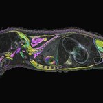
A living sea urchin larva (Arbacia punctulata) under a polychromatic polarizing microscope 🔬
Its calcite skeletal rods are birefringent. Here the hue shows the crystal's orientation, so colors shift as it swims. No stains.
#Microscopy #MarineBiology
Michael Shribak retweeted

📢The public vote for the BioTechniques Image Competition 2026 is NOW OPEN!!
This year, the submissions were so strong that there’s a three-way tie for tenth place, meaning our Top 10 is actually a Top 12! You can check out this bumper crop of gorgeous images at the link below.
Cast your vote now >>> hubs.ly/Q04y12bY0
You have until Tuesday, 6 October at 5pm BST / 12pm EDT / 9am PDT to vote. Remember, only one vote is allowed per email address recorded.

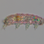
Not one of these colors is pigment.
A tardigrade under a polychromatic polarizing microscope. 40x, half speed. It's 0.2 mm long — small enough to sit inside a printed period. 🧵
Awake and walking, like here, it dies a little above human body temperature.
The toughest animal on Earth is only tough when it's asleep.
The blazing blade near the head is the stylet apparatus — calcium carbonate spears it drives into plant cells and algae. CaCO₃ is strongly birefringent, which is why it's the brightest thing in frame.
The rainbow disc behind it is the pump.
Michael Shribak retweeted

Happy #MicroscopyMonday! Take a look at this tiny marine rotifer, Brachionus manjavacas, imaged on the Orientation-Independent Differential Interference Contrast (OI-DIC) microscope, which was invented at the MBL.
Imaged by @mshribak and @grib1.

Michael Shribak retweeted
Happy #MicroscopyMonday! This live image captures a larval sea anemone (Nematostella vectensis) that refracts light like a gemstone.
The vibrant, true-color image highlights the organism’s birefringence.
Imaged by Michael Shribak (@mshribak) and Karen Echeverri.

Michael Shribak retweeted
Diatom Arachnoidiscus ehrenbergii viewed through a Polychromatic Polarization Microscope (PPM).
Imaged by Michael Shribak (@mshribak), a senior scientist at the MBL’s Bell Center.

Michael Shribak retweeted
Happy #MicroscopyMonday!
Live Northern Star Coral (Astrangia poculata) imaged using a Polychromatic Polarization Microscope (PPM).
Imaged by Michael Shribak (@mshribak) and Loretta Roberson (@Roberson_Lo) at the Bell Center of the MBL (US Patent 9,625,369).

Visualizing Biaxial Birefringence: These conoscopic interference patterns of a 25-µm DuPont HB3 biaxial film. •Left: crossed linear polarizers. •Right: Polychromatic Polarizing Module (PPM). PPM technology is available through MBL #Microscopy #Birefringence #Crystallography

Michael Shribak retweeted
This polychromatic polarizing microscope image of 0.3µ thick calcite film shows real colors, as it is seen by naked eye. The vibrant colors are produced as the calcite crystals form, revealing their molecular orientation.
#MicroscopyMonday

Super-resolution single-molecule fluorescence with quantitative phase contrast. This combo enables ~0.05 nm optical path difference detection, < 20 nm resolution, and continuous live-cell imaging with digital staining
doi.org/10.1101/2025.10.13.6…
#Microscopy #OIDIC #SMLM
Polychromatic polarizing microscope image of freezing water. The vibrant colors are produced as the ice crystals form, revealing their molecular orientation The innovative polychromatic technique directly maps slow axis orientation to color hue. DOI: 10.1080/02678292.2022.2142883

Diatom Arachnoidiscus. Diameter 220 µm. The live image taken with polychromatic polarizing microscope (researchgate.net/publication… )

Michael Shribak retweeted

My PhD students @katsu_s_minami and @shiori_iida have won the 🏅Morishima Award for their outstanding PhD research @NIG_idenken.
Huge congratulations 🎉 — I’m so proud of them! 👏👏

Mr. Minami, Katsuhiko, Ms. Iida, Shiori, and Mr. Hatoyama, Yuki received the Morishima Award
nig.ac.jp/nig/2025/03/inform…

Michael Shribak retweeted
I am seeking an Assistant Professor in my lab. If you are interested, please contact me! 📩
国立遺伝研ゲノムダイナミクス研究室で助教を公募中です。ご興味のある方はぜひご連絡ください!
RTお願いします。 🔄
#AcademicJobs #AssistantProfessor #助教公募

Michael Shribak retweeted
Exciting news! MBL has received a $2.6M gift from an anonymous donor! This generous support fuels our mission of discovery, education, and innovation. Private philanthropy drives groundbreaking research and shapes the future of science. 🌎🔬 #ScienceStartsHere #Philanthropy

ALT A drone view of the Lillie Building at the Marine Biological Laboratory