

Research Scientist at MIT, NCI K99/R00 Pathway to Independence Awardee
Cambridge, MA
Joined May 2011
- Tweets493
- Following341
- Followers385
- Likes2.7K




I've spun-out the framework used in our two-photon microscopy for cancer surgery into a set of packages called Dirigo: github.com/dirigo-developers…
While it's a work in progress, I've been trying to write weekly-ish posts on design and features: Post #1 ...

#1 Intro: forum.image.sc/t/designing-d…
#2 Demo and pipeline: forum.image.sc/t/introducing…
#3 HW extensibility: forum.image.sc/t/introducing…
#4 Unit quantities: forum.image.sc/t/introducing…

Looks interesting: <$1000 gigasample/sec software-defined oscilloscope
crowdsupply.com/eevengers/th…
Tim Weber retweeted

👀 Some of the proposed institute names are too general… National Institute on Body Systems Research? Some seem to lose the range of funded activities… NICHD to National Institute for Disability Related Research? 🤔 Others stay as is.
Full report: d1dth6e84htgma.cloudfront.ne…

A new congressional blueprint for restructuring the NIH has been released. It proposes consolidating NIH's 27 IC's and centers into 15 categories, plus term limits for directors. It adds a national security review to grant apps. Public comments due Aug. 16 energycommerce.house.gov/pos…

Tim Weber retweeted

⚡️📣👇Tremendously excited to share our new @CellCellPress article, where we develop TriPath, a method for analyzing 3D pathology samples using weakly supervised AI. Article: authors.elsevier.com/a/1j3Ri…. TriPath enables 3D computational pathology via 3D multiple instance learning allowing AI models to capture intricate morphological details from pathology volumes.
Code: github.com/mahmoodlab/TriPat…
Blog post: linkedin.com/pulse/towards-3…
Tested on two different imaging modalities, and patient cohorts from two institutions. Our superstar @GreatAndrew90 put in a monumental effort of leading the study, in a fantastic collaboration with @jonliu123 at @UW .
Interesting aspects:
- Utilizing the whole tissue volume and leveraging 3D deep learning enable superior risk prediction performance compared to 2D deep learning baselines based on a few sampled tissue sections that emulate standard clinical practice. This indicates TriPath can harness additional information provided by 3D tissue morphology.
- The performance is also superior to clinical baselines from a reader study that involved six expert pathologists.
- The morphologically heterogeneous tissue volume could lead to opposing patient-level outcome predictions, dependent on which portion of the tissue volume is used. This concurs with current clinical literature warning that tissue sampling bias can lead to misdiagnosis.
Some limitations:
- While the 3D pathology cohort size is unprecedented, it is smaller than typical 2D pathology cohorts. Further large-scale studies will be required for validation. Nevertheless, we believe that this study will initiate a positive cycle, encouraging academic institutions and pharmaceutical companies to contribute large banks of human tissue blocks with paired clinical outcomes, thus speeding up advancements in 3D computational pathology.
Concluding insights: We believe that 3D pathology is just around the corner - It has the huge potential to not only augment/improve the current clinical practice centered around 2D examination of human tissue, but also help reveal novel biomarkers for prognosis and therapeutic response..
@harvardmed @harvard_data @MassGenBrigham @broadinstitute
New preprint from @AdamEzraCohen's group shows that sensitivity may be recovered if longer wavelengths (beyond that of Ti:sapph, sadly) are used. biorxiv.org/content/10.1101/…


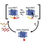
Voltron 1&2 are excellent voltage indicators, combining the sensitivity/targeting of genetically encoded microbial rhodopsin with the approaching-perfect photophysical performance of engineered organic dyes. However... (gif: doi.org/10.1038/s41593-023-0…)

This is a good idea--using an eyepiece just before the PMT.

Many years ago, I started using telescopes composed of a f=90 mm achromat and a 19 mm @TeleVueOptics Panoptic eyepiece made for amateur astronomers for the 5 mm GAsPs detectors.

Tim Weber retweeted

Our end-to-end workflow for 3D pathology is now published in @NatureProtocols!
This includes all the steps to go from archived pathology tissues to 3D H&E-like datasets, with an emphasis on quality control for large studies.
Full text at: rdcu.be/dwIYW
Would anyone be interested in such a package (light weight & free laser scanning microscopy controller written in python)? What sort of hardware compatibility would you want?

Prof. Fujimoto thoroughly enjoyed the video!


#LaskerAward winners Fujimoto, Swanson, & Huang invented optical coherence tomography, which allows doctors to rapidly detect diseases of the retina. #OCT has saved the eyesight of millions. #Lasker2023
youtu.be/UyZXJ3WjKwI
@MITEngineering @MITEECS @MIT @CaseyEye @OHSUNews


Thank you for laying the foundations with your introduction of multiplane confocal (multi-z)! opg.optica.org/optica/fullte…
This Post is from an account that no longer exists. Learn more


Kudos to @eLife for injecting some much-needed creativity to the stale institution of scientific publishing. I'm not sure this works but it's worth the try.
elifesciences.org/articles/8…