

Cell biologist at UCL. All opinions are my own!
London
Joined January 2012
- Tweets179
- Following164
- Followers447
- Likes101





My review with Chris Loewen and Will Prinz of the history of research into inter-organelle communication is out:
journals.biologists.com/jcs/…
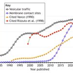
The image shows contact research rocketing upwards, and shows how a key lipid traffic paper was foundational, while a Ca2+ flux papers was not.

We have just published new research from Matthew A. Schilling and Karin M. Reinisch (Yale):
Bacterial AsmA Proteins* Transfer Glycerophospholipids Between Membranes in Vitro
journals.sagepub.com/doi/ful…
*TamB and YhdP

Thanks to James, Alyssa and the whole team for getting this story out in Nature.


1/ Thrilled to share our new paper in @Nature: “CLCC1 promotes hepatic neutral lipid flux and nuclear pore complex assembly.” From our lab together with @apsarruda, led by co–first authors @alymathiowetz and @EmilyMeymand. 🥳
nature.com/articles/s41586-0…

Also, quite coincidentally to me working at @UCLeye, a mutation in CLCC1 causes a rare autosomal recessive #Retinitis (RP32)
ncbi.nlm.nih.gov/pmc/article…
Is the molecular basis of that blinding disorder related to nuclear pore complex insertion? Or another nuclear envelope function?


Great to see our preprint out. We uncovered a simple likely mechanism to fuse inner+outer nuclear envelopes to precede nuclear pore complex (NPC) insertion.
Our first step was to show that CLCC1 is the long-lost human homologue of Brl1 (previously only known in yeast, protists)
New preprint w/ @apsarruda! Tour de force from grad student extraordinaire @alymathiowetz! >20 CRISPR screens enabled the discovery of a key role for CLCC1 in hepatic neutral lipid flux and nuclear pore complex assembly. Many surprises! #LipidDroplets 1/3 biorxiv.org/content/10.1101/…

2/3
Interestingly both CLCC1 (human) and Brl1/Brr6 (yeast) both have strong lipid droplet phenotypes too.
Next we made AF2 predictions of oligomers. Both proteins are predicted to form ring oligomers.
Here's the CLCC1 ring (yellow=TMH, pink=amphipathic, same as Brl1 ring above)
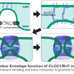
3/3 A major feature of these rings is that they deform the membrane into the lumen. Two opposing rings on inner and outer nuclear envelopes might lead to fusion, forming a pore that then opens to accommodate the NPC.
Congratulations to @alymathiowetz & the whole team.

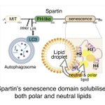
Yaoyang Zhong, PhD student in my lab, & I have a new News and Views in @ContactCTC about the discovery by Karin Reinisch's team (Yale) that the senescence domain in Spartin is a lipid transfer protein doi.org/10.1177/251525642412…
The original work is in PNAS: pnas.org/doi/10.1073/pnas.23…

Excellent new paper from the lab of Sabrina Buttner (& collaborators incl. me!) showing LDO proteins bridge from lipid droplets to the yeast vacuole to allow lipophagy during starvation.
Could the animal LDO-ologue promethin (=LDAF) act similarly? it has ~30aa disordered tail.

We are so proud to share the final version of our latest #MembraneContactSites story🎉, now in @Dev_Cell. We identify the LDO proteins and Vac8 as a tethering pair between the vacuole and lipid droplets (vCLIP) that mediates microlipophagy in starvation
cell.com/developmental-cell/…

Our lab has a new preprint out on how LAM proteins self-organise into puncta. This turns out to NOT positively affect function ... rather the opposite.
biorxiv.org/content/10.1101/…
+We collaborated with @KukulskiWanda to idenitfy the ER architecture at LAM+ puncta: it's unremakrable.

Elife is moving forward but its claim to be “relinquishing role of gatekeeper” applies only to removing gate #2.
Elife will still keep gate #1: “we do not have the capacity to review every preprint submitted to us”. Deciding what to review is now uniquely important... & secret!


Massive news: eLife to abolish accept/reject decisions: papers will just be “peer reviewed”. Others can argue about this, but lots of interesting consequences. 1/9 elifesciences.org/articles/8…

My paper about VPS13 ATG2 +3 other eukaryotic bridge-like lipid transfer protein families made from repeating beta groove (RBG) domains is now out in
@ContactCTC .
journals.sagepub.com/doi/10.…
I uses sequence homology & structural predictions to analyse the proteins
(1 of 5)

4 of 5
Viewing the most conserved residues in detail shows that there are many hydrophilic residues all the way along the outside of the lipid-binding groove, well placed to bind protein partners.


2 of 5
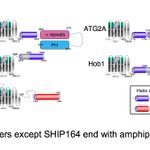
Comparing all the RBG domains in VPS13 with each other identified just four types:
• the extreme ends (x2)
• just 2 central types (here shown in blue/red) that have duplicated many times


Our 6-way collaboration is out, led by Schuldiner lab @WeizmannScience, @conibear_liz lab @UBC & Chris Stefan @LMCB_UCL. Also Vanina Zaremberg @BioSciUofc & Robert Ernst SaarlandU
We found lots of new contact site proteins, many of which transfer lipid
elifesciences.org/articles/7…

Since our new PM is not going to #cop27egypt, @Keir_Starmer should go - that would send a very strong message on leadership.
