

I am a biomedical engineer who develops new technologies for imaging the brain and retina with light
New York, NY
Joined February 2018
- Tweets75
- Following59
- Followers198
- Likes114




See the outer retina in 'full color' with visible light OCT: tinyurl.com/48j43wr3
@NYULangoneEye @NIH_CommonFund #NEIfunded (partially)

ALT Visible light OCT shows spectral properties of outer retina bands (red shift towards deeper RPE)
Vivek Srinivasan retweeted

.@nyulangone is a recipient of an Oculomics grant from the @NIH_CommonFund (partially #NEIfunded). The plan is to investigate how OCT can be used to detect neurological diseases like Parkinson’s, Alzheimer’s & more.
@vjsriniv @NYULangoneEye
More: bit.ly/3TniriY

ALT Three images showing an OCT image of an eye, and layers of the retina, CAPTION: Commercial near-infrared (NIR) and visible light Optical Coherence Tomography (OCT) of the same eye. Compared to commercial NIR OCT, visible light OCT achieves fivefold finer axial resolution, which improves visualization of the ganglion cell layer, inner plexiform layer, and inner nuclear layer.
Vivek Srinivasan retweeted

Dr. Huang co-invented an imaging technology that routinely helps prevent blindness and is increasingly used to diagnose and treat conditions of the heart, brain, skin and more. #Lasker2023 #LaskerAwards spr.ly/6010u6SD4
Visible light OCT can now image macular pigments (MPs), potent absorbers of harmful blue light. Read about the first localization of MPs to Henle's fiber layer in the living human retina! #OSA_OL @NYULangoneEye #aging
opg.optica.org/ol/abstract.c…

Vivek Srinivasan retweeted

A team led by @NYULangoneEye's Dr. Vivek J. Srinivasan (@vjsriniv) used visible light OCT to gain new insights into retinal regions such as band 2 that can ultimately help improve diagnostic capabilities and treatments for retinal disorders.
Read more: bit.ly/3pQ8hwk

Vivek Srinivasan retweeted

Authors report imaging rod photoreceptors in the aging mouse eye from soma to synapse for the first time. Also, authors find that visible light Optical Coherence Tomography uncovers yet another retinal band. ow.ly/jGOl50Oi6vS @NYULangoneEye @ch21pooja @vjsriniv

Yet another retinal band uncovered by @ch21pooja and @AaronKho4 in the living eye with visible light OCT! Now six new retinal features...and counting. Read here about our latest discovery on rod spherule synapses: tinyurl.com/28xbujhv
@NYULangoneEye @nyugrossman @AAOjournal
Vivek Srinivasan retweeted

Ph.D. fellowship - We are seeking a doctoral student to join our research group at @UniStavanger. We are interested in developing bacterial polymeric biomaterials for biomedical applications.
RTs would be highly appreciated :)
Here is the advertisement
jobbnorge.no/en/available-jo…
Vivek Srinivasan retweeted
SPIE @PhotonicsWest Ophthalmic Technologies:
Really nice work in ocular imaging methods & applications. Many #NEIfunded, including:
@ojsaeedi @YaliJiaOCT @vjsriniv @rjonnal @IU_Optometry @dukeeyecenter @SabesanLab @OSUOptometry @schalleklab @ucd_eyecenter @uicovs #PhotonicsWest

ALT Photo of audience at SPIE Photonics West Conference (Ophthalmic Technologies session)
Vivek Srinivasan retweeted

Glenn Yiu et al. @ucd_eyecenter @NatEyeInstitute @bcmhouston characterized a primate model of myopic foveoschisis by clinical examination & imaging followed by morphologic, cellular, and transcriptional profiling of retinal tissues and genetic analysis.
doi.org/10.1167/iovs.64.1.18

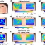
Just out in Optica...human #brain #opticalimaging with a highly parallel, time-of-flight informed approach! Interferometric #NIRS charges ahead! tinyurl.com/mupvc2rn
@NYUGrossman @ucdavisNCIBT @OpticaWorldwide

Outlook on the future of interferometric diffuse optics: dx.doi.org/10.1117/1.NPh.10.….
We look forward to disseminating these exciting technologies to the community through @ucdavisNCIBT!

Vivek Srinivasan retweeted

Rolling out a new preprint on 2nd Gen dual-channel visible light optical coherence tomography. 👇👇biorxiv.org/content/10.1101/… Several years in making, too. We learned first hand balancing resolution and imaging range is challenging in clinics. We proposed a solution for it.


Vivek Srinivasan retweeted
Largest clinical study so far with visible light OCT. Several years in making, and perhaps the most challenging project that I have had so far, technically and practically. Shout out to @WeiyeSong and all authors! Scattering Spectroscopy in the human eye has bright future!

Ji Yi et al. @The_BMC find that visible light OCT of peripapillary retinal nerve fiber layer reflectivity better distinguishes suspect/preperimetric glaucoma eyes from normal eyes than Cirrus OCT thickness measurements.
doi.org/10.1167/tvst.11.9.28

Vivek Srinivasan retweeted

The NCIBT kick off symposium was a great success! Thank you every one who contributed and participated in the stimulating discussions. @UCDavisHealth @UCDavisCOE @carney_lab @MarcuLab @vjsriniv @Leibniz_IPHT @Brainlab @saifislam @KwanliuMa @AijunWangLab and many more

Vivek Srinivasan retweeted

Visible Light Optical Coherence Tomography Reveals the Relationship of the Myoid and Ellipsoid to Band 2 in Humans ift.tt/ThEFRu6
Just out in @ARVOtvst-More subcellular detail revealed by visible light OCT: myoid-ellipsoid boundary and band 2a for cones as well as band 2b for rods! What will we find next?
Read here: doi.org/10.1167/tvst.11.9.3
@NYULangoneEye @NYUImaging @UCDavisBME
Vivek Srinivasan retweeted

This #neurophotonics issue was one of the hardest to coordinate but the most rewarding, too! The result is one of the most complete references in our #fnirs field. I wanted to thank everyone who contributed and hope you all enjoy reading it as much as I enjoyed working on it!
New visible light OCT retinal bands, imaged in the living eye and validated by histology! Stay tuned...more new bands to come. @NYULangoneEye @NYUImaging @UCDavisCOE doi.org/10.1167/iovs.63.9.10


Vivek J. Srinivasan @vjsriniv et al. @UCDavisCOE
combine in vivo, 1.0-µm axial resolution visible-light OCT and ex vivo electron microscopy to investigating three subcellular features in the mouse outer retina.
doi.org/10.1167/iovs.63.9.10
