

Epileptologist. Epilepsy fellowship director. Teaching EEG and epilepsy, building EEG learning tools. Opinions are my own.
Detroit, MI
Joined April 2023
- Tweets79
- Following289
- Followers202
- Likes572





Pinned Tweet
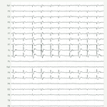
LPDs +F in an ICU patient.
Which hemisphere is the source in?
Image 1: average reference. Image 2: bipolar.
The answer and a 3D movie of the source are in the reply.
Synthetic EEG, educational use only. eegsimlab.org #EEG #MedEd


This weekend I read critical care EEGs with seizure and spike detectors enabled. You would think that makes the work easier. With the detectors currently available, it does not!
I think a core problem is treating EEG as a pattern recognition problem. It is true that we spend hundreds of hours training our own neural networks to pick up the patterns, but that is only one layer in a reasoning system. That system lets us compare waveforms, drop or raise our own detection thresholds, and incorporate data from video and charts to reach a diagnosis.
What we need are detectors reliable enough to take over the pattern recognition, and built to feed into how we actually reason through a record.
43
Part of EEG training is learning to ignore the sea of noise and pick up the 1% that matters.
Early in training, I was overwhelmed by all the EEG data I had to review, until I learned that most is clinically irrelevant. Knowing what to ignore is as important as picking up what matters.
In the bipolar image:
Right: Fp2-F4 down, F4-C4 flat, C4-P4 up. F4 and C4 are equally negative.
Midline: Fz-Cz flat, Cz-Pz up. Fz and Cz are equally negative.
Left: small deflections only.
A source on the medial surface can project its field to the other side.
1
36
Answer: LEFT.
The source is a patch of cortex on the left medial frontal surface (pre-SMA). It faces up and to the right.
So the negative field lands on Fz, Cz, F4 and C4, and the left side goes slightly positive.
The EEG looks right-sided. The source is on the left.
1
34
Waleed R Abood retweeted

Join us for the 5th International Conference on #AI in #Epilepsy & Neurological Disorders (February 22–24, 2027), followed by the SEEG Research Update Symposium (February 25, 2027), in Breckenridge, Colorado!
aiepilepsy-neuro.com/

Aplying to epilepsy fellowship? Here is a 2-minute look at our program.
youtu.be/x9zB0ZkWoSM
At Wayne State University and Detroit Medical Center, fellows train at a Level 4 epilepsy center and may choose an adult or pediatric focus.
Highlights:
• More than 1,000 EMU admissions in 2025, including stereo-EEG
• Epilepsy surgery: stereo-EEG, laser ablation, RNS and VNS
• Advanced EEG: EEG source imaging and rapid-response EEG
• Eleven adult and pediatric faculty, with a weekly conference and weekly didactics
• No overnight call
We accept two fellows each year through ERAS and NRMP.
#Epilepsy #Neurology #MedicalEducation #Fellowship
We just released two new eegsimlab.org lessons:
• What is an EEG? eegsimlab.org/whatis
• What is a montage? eegsimlab.org/montage
Also new: dark mode and a tablet/laptop view.
#EEGSimLab #EEG #MedEd

Next up: EEG technology, how to read an EEG, how to write a report, and the EEG of the epilepsy syndromes.
Tried a lesson? Tell us what to fix with the Feedback button on eegsimlab.org.
Educational use only, not for clinical care.
2
225
Awake EEG. Which dipole made these spikes?
Image 1: bipolar EEG. Image 2: four dipoles on the left side of the head.
Reply A, B, C or D.
Synthetic EEG, educational use only. #EEG #MedEd
eegsimlab.org


Move the dipole yourself:
eegsimlab.org/dipole?utm_sou…
4
251
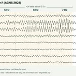
What is this run?
A left temporal run builds from 5 to 6 to 7 Hz, grows in voltage, and stops after about 6.5 s.
Vote in the poll below.
Synthetic EEG, educational use only. eegsimlab.org #EEG #MedEd

1
9
1,359
EEGSimLab is now built for your phone: ACNS 2021 EEG terms with key numbers, guided lessons you swipe through, IFCN spike criteria and a one-dipole demo. Synthetic EEG, free, educational use only. eegsimlab.org #EEG #MedEd
🤖 Made with AI
A note on EEGSimLab: the code was built entirely with Claude Code. I designed and verified the teaching content. It’s descriptive: synthetic EEG drawn to show what each term means, not to look like real recordings. Educational use only. eegsimlab.org
1
3
231
5/ Settings > Capabilities > Allowed websites. Add:
Required:
eutils.ncbi.nlm.nih.gov (PubMed search)
ebi.ac.uk (Europe PMC: papers and open full text)
clinicaltrials.gov (trial registry)
api.crossref.org (checks DOIs and retractions)
Recommended:
api.openalex.org (finds citing and cited papers)
api.datacite.org (checks DOIs Crossref doesn't hold)
doi.org (confirms DOI links work)
All free public services. The skill only reads from them.
1
27