

ECG ❤️ ECHO beginner
Srbija
Joined February 2022
- Tweets1.4K
- Following855
- Followers395
- Likes2.3K





Ivan Bušić retweeted

Uhl’s anomaly in a pediatric patient
📌 virtually absent RV myocardium
📌 paper-thin RV wall
📌 severely dilated RA and RV
Rare, congenital - and not exclusively pediatric. Adults can present with it too.
@iamritu @OungSavly @DrRajeshG1 @CASivaram1 @loomba_rohit

One dilated RV. Three possibilities. 👀.
How do you tell them apart on echo? 🫀
doi.org/10.1111/echo.13430
Ivan Bušić retweeted

Let's repeat how the infranodal AV block improves upon slow-down of P waves. nitter.cf/KostekMilan/status/185…


Ivan Bušić retweeted
Multifocal Ectopic Purkinje-related Premature Contractions (MEPPC) Syndrome
sciencedirect.com/science/ar…

Ivan Bušić retweeted
What is DOT-CRT ❓
LOT-CRT patients were subclassified into “successful LOT-CRT” (LBBAP; presence of r’ in electrode electrocardiography [ECG] V1) and “deep septal optimized therapy” (DOT-CRT) (functional deep septal capture).

Echocardiographic response from Left Bundle Branch Area Pacing Optimized Cardiac Resynchronization Therapy (LOT-CRT) versus traditional CRT #OpenAccess
@pvijayaraman1
heartrhythmjournal.com/artic…


Ivan Bušić retweeted

Understanding LV Diastolic Dysfunction via Mitral Inflow Doppler:
Mitral inflow Doppler patterns reflect LV diastolic function. Pulsed-wave Doppler at the mitral valve leaflets shows E (early filling) and A (atrial contraction) waves.
Here’s how the patterns change:
1️⃣ Normal Filling:
- E/A >1
- Deceleration Time (DT) <220 ms
- Normal relaxation and compliance.
2️⃣ Grade I – Mild Dysfunction (Impaired Relaxation):
- E/A <1
- DT >220 ms
- Relaxation is delayed, atrial kick becomes dominant.
3️⃣ Grade II – Moderate Dysfunction (Pseudonormal):
- E/A >1 (appears normal!)
- DT 150–200 ms
- LA pressure increases to "normalize" filling—requires Tissue Doppler to unmask.
4️⃣Grade III – Severe Dysfunction (Restrictive Pattern):
- E/A >1.5
- DT <150 ms
- Very high LA pressures, stiff LV—hallmark of poor prognosis.
⚠️Always correlate with tissue Doppler (E/e’), LA size, pulmonary vein flow, and clinical picture. A pseudonormal pattern can be misleading without these.
Ref:
Ho CY, Bulwer BE. In: Solomon SD, Bulwer BE. Essential Echocardiography. Humana Press, 2007:124.
#CardioTwitter #EchoFirst #MedX


Ivan Bušić retweeted
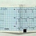
PAC or PVC? Only the chest leads. A moment's thought should suffice to answer the question.

Ivan Bušić retweeted

#Sharing is learning
An interesting finding on the #Aortic #Valve in a patient with #Mitral #Stenosis. There are no #symptoms or #history of #infective #endocarditis.❗ F, 56 y.o @Irina67790690

Ivan Bušić retweeted
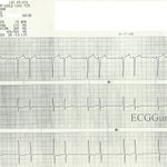
Many signs in this ECG indicate ventricular tachycardia. Name the signs that rule out supraventricular tachycardia with aberrant conduction.

Ivan Bušić retweeted

🎥 Apical Rocking
🔍 Classic echo finding in LBBB
🫀 Caused by septo-lateral dyssynchrony
💡 Key marker of mechanical dyssynchrony → may predict CRT response
👉 Always watch the apex carefully!
#Cardiology #Echocardiography


Ivan Bušić retweeted

Amazing #ECG ✅
#ESCCongress2025 #ESCCongressVisuals 🇪🇸
👇
@ecgandrhythmRoe
@ECGEPSCADEVICE
@ecgrhythms
@OrlanditoCardio
@raico980724


Ivan Bušić retweeted

🫀 #CardioNugget
The “Lobster Claw” Sign 🦞 in HOCM
In HOCM, the LV outflow tract is narrowed mid-systole due to systolic anterior motion (SAM) of the mitral valve + septal hypertrophy.
📉 This leads to a bimodal LV ejection velocity pattern on Doppler—initial acceleration, followed by a mid-systolic drop (from dynamic obstruction), then a late rise.
🎯 The result? A “lobster claw” or “spike and dome” contour—diagnostic of dynamic obstruction!
🧠 Pro tip: Seen best with continuous wave Doppler across the LVOT. If you see a claw, think obstruction!
#CardioNuggets #MedEd #CardioTwitter

Ivan Bušić retweeted

Significant Pulmonary Regurgitation.
10, y.o.
Нas a history of open pulmonary valve valvuloplasty 5 years ago.

Ivan Bušić retweeted

👉 Typical vs. Atypical AVNRT – Mechanistic Overview 👈
👉 Typical AVNRT (slow–fast):
Impulse conduction: Atrium → Slow Pathway → Ventricle → Fast Pathway (retrograde)
•This produces a short RP tachycardia, because the retrograde atrial activation from the ventricle back to the atrium occurs via the fast pathway.
•Consequently, the P wave is inscribed at the terminal part of the QRS complex on the ECG.
👉 Atypical AVNRT (fast–slow):
Impulse conduction: Atrium → Fast Pathway → Ventricle → Slow Pathway (retrograde)
•This produces a long RP tachycardia, because the retrograde atrial activation from the ventricle back to the atrium occurs via the slow pathway.
•Therefore, the P wave appears after the QRS complex on the ECG.
#EPeeps This post was created in collaboration @N_Trajkovska
@Phiso_de @chris_sohns @MBraunEP @MoneebKhalaph @Cardioschool @GNageler @ecgandrhythmRoe @Ecgloverr @ecgrhythms @KostekMilan @ALFIEEP1 @ECGEPSCADEVICE @nelsonpol @pjsm83 @ChristianHeeger @narrowQRS @MeldaYesilyurt @JNJNews @AbbottNews @BSCCardiology #CardioTwitter @TobiasReichlin @ArashArya_EP
Ivan Bušić retweeted
Doming PV in a pediatric patient with bicuspid pulmonary valve (BPV) 👉 UE Ao Arch SAX #echofirst
